EMG BioInputs
This project has not been finished yet, stay tuned for the next release.
Introduction
Electromyography (EMG) is a widely-used technique for recording and analyzing electrical activities generated by skeletal muscles.
EMG is appealing due to its simplicity when detecting basic movements, such as a biceps contraction. For such projects, advanced hardware or complex signal processing is often unnecessary. This makes EMG an accessible tool for beginners as an introduction into biomedical engineering projects. Furthermore, the foundational concepts learned through EMG can be extended to related fields, including Electrocardiography (ECG), Electrooculography (EOG), Electroencephalography (EEG), and others.
In SensEdu, we demonstrate how basic biceps contraction can be used to simulate keyboard press logic, with latency of ~100–200ms. We use the most basic circuit design combined with signal processing in MATLAB.
This page will guide you step-by-step through the required setup and knowledge to create a simple yet effective demonstration of a typical biomedical engineering project.
Biological Background
First, we need to establish an understanding of what we are trying to measure.
Highly recommended watching the video Neuromuscular Junction by Byte Size Med.
The most important structure that we need to focus on is the Neuromuscular Junction (NMJ). This is a specialized synapse that acts as a communication bridge between motor neurons and skeletal muscle fibers. The NMJ plays a fundamental role in converting neural signals into muscle contraction. Let’s break down its working principle step by step.
The process begins with a motor neuron transmitting an electrical signal called an Action Potential. This signal travels along neuron’s axon until it reaches the NMJ. It activates voltage-gated sodium channels, which allow positively charged sodium ions (Na⁺) enter the cell. The influx of sodium causes the cell membrane to become “less negatively charged”, a change known as depolarization.
Continuous depolarization of the cell membrane subsequently activates voltage-gated calcium channels. Calcium ions (Ca²⁺) flow into the cell which triggers little containers with neurotransmitters to fuse with the membrane and release Acetylcholine (ACh).
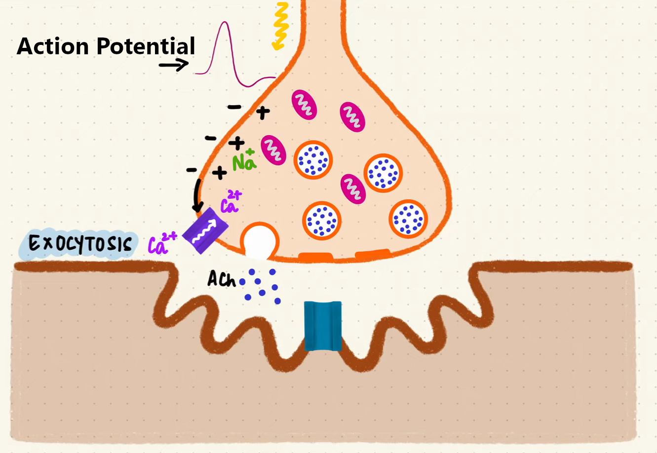
Exocytosis process at the Neuromuscular Junction (source: Byte Size Med)
Acetylcholine travels across the synaptic space and binds to specialized receptors on the muscle cell’s membrane. These receptors, also known as ligand-gated ion channels, respond to the chemical messenger (neurotransmitter) by opening and allowing ions to enter the muscle cell. This process triggers membrane depolarization, causing the resting membrane potential of the muscle cell (normally at around -90mV) to become less negative.
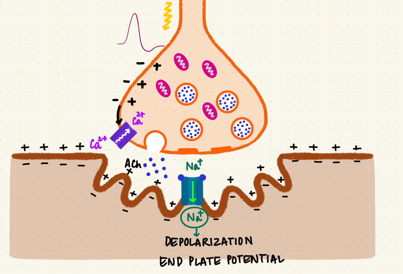
Depolarization caused by the opening of ligand-gated ion channels (source: Byte Size Med)
As more ACh molecules bind to their receptors, the membrane voltage continues to rise. Once it reaches a threshold of approximately -50 mV, voltage-gated sodium channels open, allowing sodium ions (Na⁺) to rush into the muscle cell. This influx of sodium completes the process of membrane depolarization and generates an Action Potential. This electrical signal propagates along the muscle membrane, eventually leading to a muscle contraction.
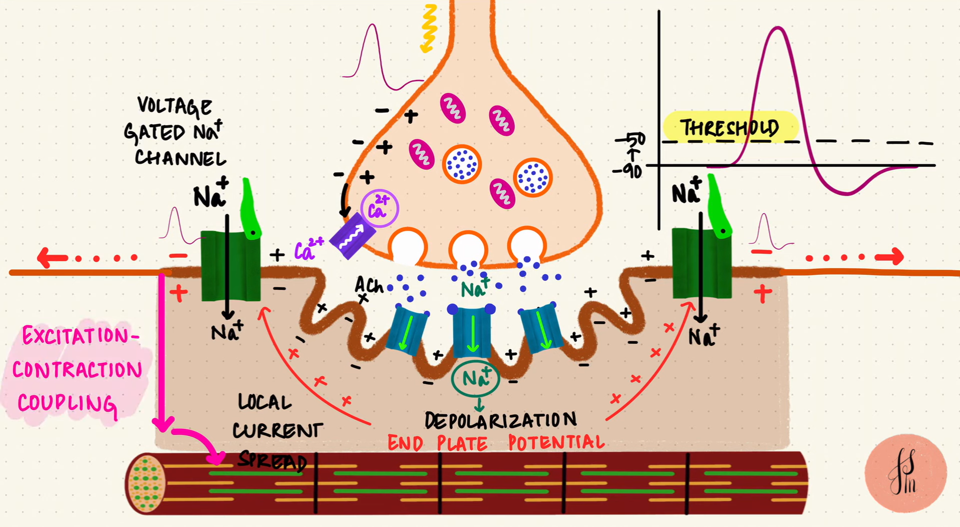
Excitation-Contraction Coupling (source: Byte Size Med)
If you feel overwhelmed by the details, the main takeaway: a neural electrical signal is converted to a chemical signal and then back into electrical signal in the muscle. The sequence Neural Action Potential → Acetylcholine Release → Muscle Action Potential. The muscle action potential then propagates along the muscle cell membrane and ultimately triggers muscle contraction.
The most important point is that this muscle action potential can be measured, providing valuable information about muscle activity.
Recording Action Potentials
Action Potentials can be recorded using different methods:
- Invasive Needle EMG: Inserting electrodes directly into the muscle, enabling highly precise and localized recordings. It is widely used in clinical applications and treatments where precision and measurement quality are critical.
- Non-invasive Surface EMG: Placing electrodes on the skin above the muscle. This approach improves patient comfort and simplifies repeatable studies, making it ideal for educational purposes. However, sEMG has significant precision limitations due to the layers of skin and fat between the electrodes and the muscle. These limitations make it challenging to differentiate the activity of individual muscle fibers and to record signals from small muscles, like ones around the fingers.
In SensEdu we use surface EMG. From now on, whenever we mention EMG or electrodes, we are referring to sEMG and surface electrodes, respectively.
Hardware Setup
This section describes the necessary connections and hardware details required for the proper setup of the EMG BioInputs system and writing the script for MCU. Below is an illustration of the overall system architecture.
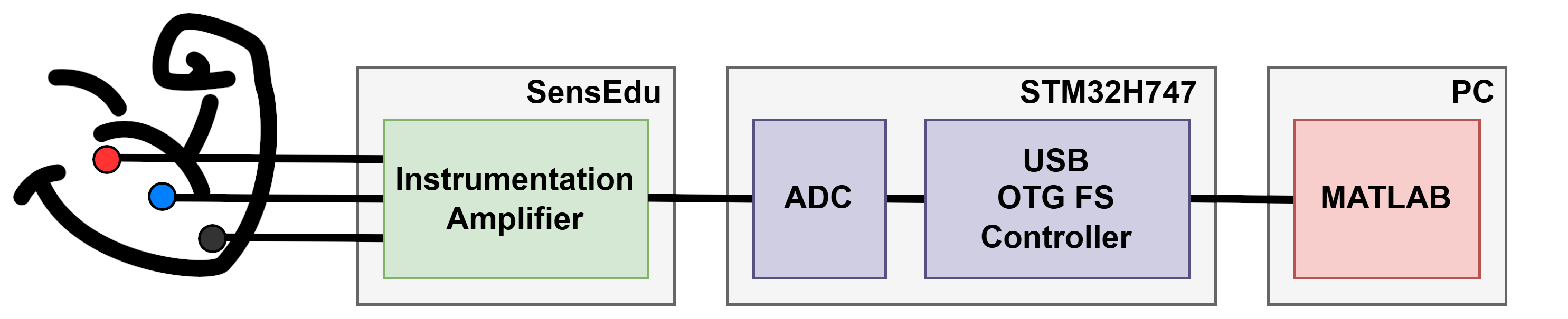
EMG BioInputs System Architecture
Electrodes Connectivity
Under construction.